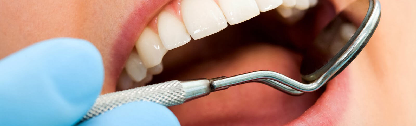
Procedures and Patient Information
Gingival Grafting
What causes gingival recession?
- Abrasive and trauamatic toothbrushing habits
- Periodontal inflammation
- Tooth positioned too buccal
- Frenal and muscle attachment encroachment
- Orthodontic movement
- Underlying bony dehiscence
- Genetics
- Invasion of biologic width
What are the endpoints of success with gingival grafting?
- Gingival augmentation
- Prevent/stop recession
- Facilitate plaque control
- Elimination of an aberrant frenum
- Increasing vestibular depth
- Decreasing rot sensitivity
- Decreasing inflammation
What are some side effects from gingival therapy?
- Increase bleeding
- Bone exposure
- Color discrepancies
- Necrosis of donor site
- Herpetic lesions
- Delayed healing
What is the prevalence of gingival recession?
According to a study by Albandar and Kingman,5 prevalence of ≥ 1 mm recession is 58% in individuals 30 years and older and increases with age. Prevalence
of recession is 37.8% in those aged 30 to 39 years and increased to 90.4% in persons aged 80 to 90 years.
Gum recession treatment options include deep cleanings, pocket depth reduction, or laser gum surgery to first treat gum disease. Once your gum disease is under control, Dr.aTermeie may then recommend soft tissue grafting, or tissue regeneration to restore lost gum tissue.
Crown Lengthening
What are the indications for crown lengthening?
- Subgingival fracture/caries
- Endodontic pin/post perforation
- Root resorption
- Inadequate height for the restoration
- Esthetically short crown
- Altered passive eruption
- Biologic width would be violated during a restorative procedure
What is excessive gingival display and why does it occur?
Excessive gingival display is a condition in which a significant amount of gingiva is visible when the patient smiles. It may be caused by:
- Failure of the gingiva to migrate apically after eruption of the tooth
- Vertical maxillary excess
- Upper lip incompetence
Regeneration
What is guided tissue regeneration (GTR)?
It is the use of barrier membranes to direct the growth of new bone. It is used during wound healing.
What is guided bone regeneration?
It is similar to guided tissue regeneration. A bone graft and membrane are placed in the area of regeneration into which new bone will form.
Describe bone grafting.
Bone grafting involves replacing or augmenting missing bone around the teeth and in any areas where a tooth has been lost. The three types of bone graft procedures include: autogenous, allograft, and xenograft.
Autogenous grafts involve taking bone from one area of the patient’s body and transplanting it to the location of the mouth that needs to be restored. The bone may be taken from areas such as the chin and since the bone comes from the patient, it is still considered to be ‘live.’
Allografts are performed using cadaver bone that comes from a bone bank. The material is screened for safety to ensure optimal results.
Xenografts involve placing bone that comes from a nonhuman source such as cow, or bovine. These are typically performed when a significant amount of bone is needed for dental implants.
Is there a difference between resorbable and nonresorbable membranes for bone regeneration?
Resorbable
- No need to be removed
- Collagen membrane broken down by collagenases
Nonresorbable
- First to be developed
- Second surgery is needed to remove it
What is socket grafting or ridge preservation?
After extraction of the tooth, it is important to maintain the dimensions of the ridge. Placing a bone graft into the socket allows for this preservation especially in the esthetic zone.
Surgical Therapy
What is the primary indication for periodontal flap surgery?
The primary indication is to gain access to the root surface and remove bacterial deposits and calculus that may have remained following non-surgical therapy.
What is osseous surgery? What are the indications for osseous surgery?
It is defined as the procedures to modify bone support altered by periodontal disease either by reshaping the alveolar process to achieve physiologic form, without removal of alveolar bone or by the removal of some alveolar bone, thus changing the position of crestal bone relative to the tooth root.
Indications for osseous surgery
- Failure from treatment with gingivectomy
- Mesial of tipped molars
- Deep isolated pockets
- Deep buccal and lingual pockets
- Saucer shaped interproximal pockets
What is the rationale for osseous surgery?
- Establish access for root debridement
- Harmonious topology (positive architecture)
- Decrease pocket depth
- Preserve the teeth
- Allow for plaque control
- Improve prognosis
What are contraindications to osseous surgery?
- Proper follow-up and maintenance is not possible
- Advanced bone loss (long-term prognosis and value of the tooth is questionable)
- 3-wall defect (may be able to regenerate)
- Poor oral hygiene
- Medically contraindicated
Describe the healing following surgery
- 24 hours- There is a connection between the flap and the tooth or bone surface with a blood clot.
- 1-3 days- The space between the flap and tooth or bone is thinner and epithelial cells migrate to the site at a rate of .5 mm per day.
- One week- Epithelial attachment to the root occurs and the blood clot is replaced by granulation tissue by hemidesmosomes.
- 2 weeks- Collagen fibers form parallel to the tooth surface
- One month-Fully epithelialized gingival crevice
What are the indications for root resection?
- Severe vertical bone loss on the root not amenable to regeneration
- Furcation invasion not maintainable
- Endodontics impossible on the root
- Periodontically involved abutment tooth with a hopeless prognosis associated with a root
- Vertical/horizontal root fracture of the root.
What are the contraindications for root resection?
- Roots are fused
- Advanced bone loss with unfavorable crown/root ratio
- The remaining root on the tooth would be inadequate to serve as an abutment
- The remaining tooth would not be able to withstand normal occlusal forces
- Inadequate hygiene
What is the post-infection rate following periodontal surgery?
Powell (2005) did a retrospective study on 395 patients and included 1,053 fully documented surgical procedures. Surgical techniques reviewed included osseous resective surgery, flap curettage, distal wedge procedures, gingivectomy, root resection, guided tissue regeneration, dental implant surgery, epithelialized free soft tissue autografts, subepithelial connective tissue autografts, coronally positioned flaps, sinus augmentations, and ridge preservation or augmentation procedures. Infection was defined as increasing and progressive swelling with the presence of suppuration. He found an infection rate of 2.09% (22/1053)
How much blood loss occurs in periodontal surgery?
Zigdon (2012) did a study on Twenty-six patients who needed open flap debridement or regeneration. The amount of blood lost during the procedure ranged from 6.0 to 145.1 mL, with an overall mean of 59.47 ± 38.2 mL. Local anesthetic amount, surgical field size, and the operation duration did not relate to blood-loss volume. He concluded that blood loss during periodontal surgery is minimal.
Implant Placement
What are the criteria of success for dental implants?
- The implant is immobile (less than 1mm in any direction) and functioning
- Radiographs show no radiolucency
- Vertical bone loss is less than .2mm annually following the implant’s 1st year of service
- Implant has an absence of persistent and/or irreversible signs
- Patient and clinician are satisfied
- The hard and soft tissues are healthy
- Trained and experienced surgeon
- Maintenance and oral hygiene
- Management of the soft tissues
- Well-designed restoration
Bone Shape
- The Alveolar ridge is intact
- Moderate Ridge Resorption has occured
- Advanced Risidual ridge resorption has occured
- Some resorption of the basal bone has initiated
- Well-designed restoration
Bone Quality
- Entire Jaw is cortical
- Thick cortical bone surrounds a core of dense trabecular bone
- Thin layer of cortical bone surrounds a core of dense trabecular bone
- Thin layer of cortical bone surrounds low density trabecular bone
How much remodeling can be expected after an implant is placed?
Adell (1981)iv found that during healing and the first year after connection of the bridge, the mean value for marginal bone loss was 1.5 mm. Thereafter only 0.1 mm was lost annually.
What are some factors that affect implant success?
- Bone quantity and quality
- Surgical technique (ex: overheating of the bone and torque forces)
- Implant length and width
- Smoking
- Amount of plaque
- Occlusion (avoid: non-axial forces, cantilevers, and lateral excursives)
- Lack of Keratinized attached gingiva (KAG)
- Anatomy (lingual concavity, mental nerve, sinus location)
What factors lead to implant failure?
- Infection (Bacterial contamination and increased inflammation)
- Fracture
- Impaired healing
- Improper implant preparation
- Improper mechanical stability following insertion of the implant (mobility)
- Premature loading of the implant
- Pain
What pre-surgical steps need to be taken before implant placement?
- Restorative requirements, interarch space and jaw relationships, location of edentulous areas, and the quantity and quality of available bone should also be evaluated before implants are selected as a treatment option. It is also a good idea to get study models and photographs to be able to study the case when the patient is not present.
- Radiographs such as cone beam images or newtom scans are necessary to determine the height of available bone in three dimensions and for selection of the dimensions of the implants. They also may be needed to determine the location of anatomical structures (nerves and vessels).
- Lastly, written informed consent is discussed between the surgeon and the patient.
What is the difference between the one-stage and two-stage technique?
One-stage
The implant is exposed with a healing abutment or restoration (most likely temporary) but is not loaded.
Two-stage
The implant is placed and covered with a cover-screw and the gingiva. The patient waits 3-6 months for exposure of the implant with a healing abutment and subsequently the restoration.
What are the contraindications for immediate implant placement in the esthetic zone?
- Heavy smoker
- Patient with very high expectations
- Immunocompromised
- Highly scalloped and thin biotype
- Triangular crowns
- Soft tissue defects
- High lip line
What is platform switching?
It is the use of an abutment that is smaller than the implant. The theory is that the micro-gap is reduced and there is less bone loss.
What is the rate of failure for implants in smokers?
The reasons behind the high failure rate of implants in smokers include the following:
- Carbon monoxide reduces oxygenation of the healing tissues
- Nicotine is vasoconstrictive which increases platelet aggregation and adhesiveness and reduces blood flow
- Cytotoxic effects on fibroblasts and polymorphonuclear cells disrupt cell repair and defense
- Wound healing is impaired leading to a higher complication rate
What is peri-implantitis?
Peri-implantitis is defined as an inflammatory process affecting the tissue around an implant in function that has resulted in loss of supporting bone. Anaerobic bacteria are the primary factor for bone loss and have been observed in the sulcus of implants, especially when probing depths are greater than 5 mm.
What can cause peri-implantitis?
- Cement trapped below the gingiva
- Inadequate seating of the restoration on the abutment
- Over-contouring the restoration
- Implant malpositioning
Non-surgical Therapy
What treatment does non-surgical therapy include?
- Plaque control
- Supra and subgingival scaling and root planing (SC/RP)
- Use of chemical agents
- Oral hygiene instruction
- Recontouring defective restorations
- Occlusal therapy
What is scaling?
Scaling is instrumentation of the crown and root surfaces of the teeth to remove plaque, calculus, and staining from the teeth.
What is root planing?
A treatment procedure to remove cementum or surface dentin that is rough impregnated with calculus or contaminated with toxins or micro-organisms.
What are the end-points to successful root planing therapy?
- Excellent oral hygiene
- Pink and firm gingival tissue
- Pocket depth less than 5mmNo Bleeding on probing (BOP)
- No Bleeding on probing (BOP)
- No calculus detected
- Decreased mobility of the teeth
How effective is brushing and flossing?
Graves (1989) found that brushing decreases BOP by 35% but additional flossing decreased BOP by 67%. Floss is the most widely used method of interdental cleaning and the American Dental Association reports that up to 80% of interdental plaque may be removed by this method, resulting in a significantly reduced incidence of caries and prevention of periodontal disease
What is the Bass technique?
The bristles of a toothbrush are placed at 45 degrees to the tooth surface at the gingival margin. The bristles of the brush are moved in a back and forth motion.
Is there a difference between the types of floss and their efficacy?
Carr (2000) tested 24 dental hygiene students with four different floss types: waxed, unwaxed, woven and shred-resistant. He found minimal differences in the efficacy of different types of floss, their degree of comfort and ease of use.
Is there a difference between an electric and manual brush?
Robinson (2005) did a meta-analysis and found that powered toothbrushes
with a rotation oscillation action reduce plaque and gingivitis more than
manual toothbrushing.
Deery (2004) found no evidence of a statistically significant difference
between powered and manual brushes. However, rotation oscillation powered
brushes significantly reduce plaque and gingivitis in both the short and
long-term
How often should a toothbrush be replaced?
Glaze (1986) found that after 10 weeks, the subjects using the same toothbrush for the whole period had significantly more plaque than those who replaced their brushes every 2 weeks. As brushes deteriorated, they became less effective. No differences in gingival state were detected.
When is it an appropriate time to re-evaluate a patient following scaling and root planing?
If a patient is re-evaluated too soon, overtreatment may occur, but if a patient if re-evaluated after too long a period, the disease might progress. The optimal frequency is 4-8 weeks.
What are the endpoints to successful maintenance therapy?
- Prevent recurrence in patients who have been previously treated for gingivitis, periodontitis, and peri-implantitis
- Prevent the incidence of tooth or implant loss
- Increase the probability of locating and treating other diseases
What is the optimal frequency for maintenance visits?
Patients without additional attachment loss can be maintained every 6 months. However, most studies have reported that patients who have had a history of periodontal disease, need to be maintained at least four times per year.
What are some signs of disease recurrence?
- Recurrence of BOP
- Increased probing depth
- Radiographic bone loss
- Progressive mobility